Expert Imaging by Dr. Nikita Nemade
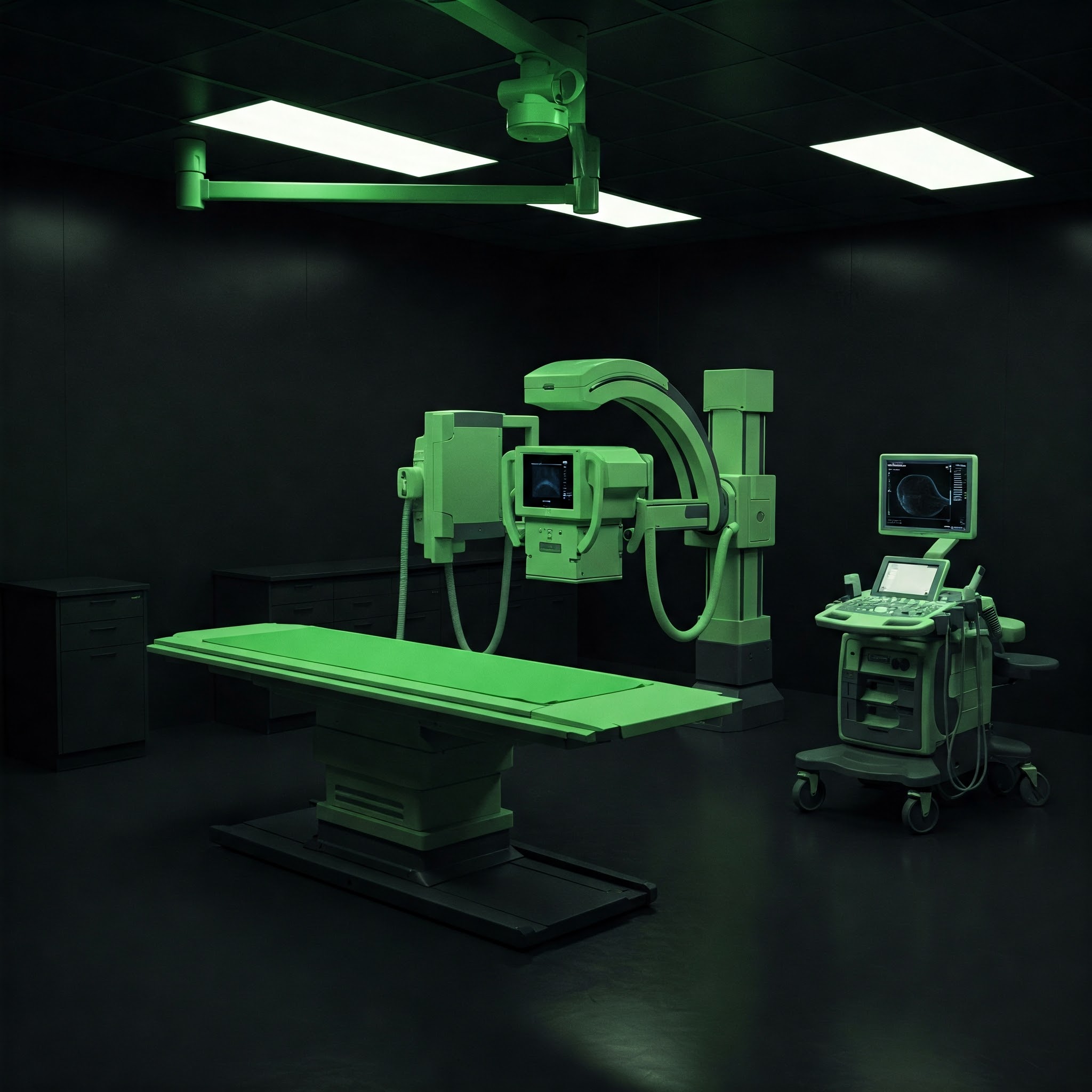
Welcome to Poorti Advanced Sonography Centre, led by Dr. Nikita Nemade, Consultant Radiologist (M.B.B.S - K.E.M. Hospital, Mumbai; MD Radiology - Pune). We provide high-quality, patient-focused sonography and digital X-ray services using state-of-the-art technology in Nashik.
Why Choose Us?
- - Expert & Caring Female Radiologist: Services overseen and interpreted by Dr. Nikita Nemade, a female doctor known for her patient-oriented approach, ensuring high diagnostic accuracy and compassionate care.
- - Advanced High-Resolution Technology: We utilize modern, state-of-the-art sonography and digital X-ray equipment, ensuring exceptionally clear and detailed images for the most precise diagnostic insights.
- - Comprehensive Services: Wide range of Ultrasound, Doppler, X-ray, and guided procedures available.
- - Patient Satisfaction & Comfort: A focus on ensuring patient satisfaction by providing a comfortable and reassuring environment throughout your scan.
- - Convenient Location: Easily accessible clinic at Mumbai Naka, Nashik, immediately next to the bus stand and auto rickshaw station. Stretcher lifts are available too.
We are committed to providing reliable diagnostic insights to support your health journey. Trust us for accurate imaging and compassionate care.
Explore our detailed diagnostic services or contact us to schedule your appointment.
Our Diagnostic Services
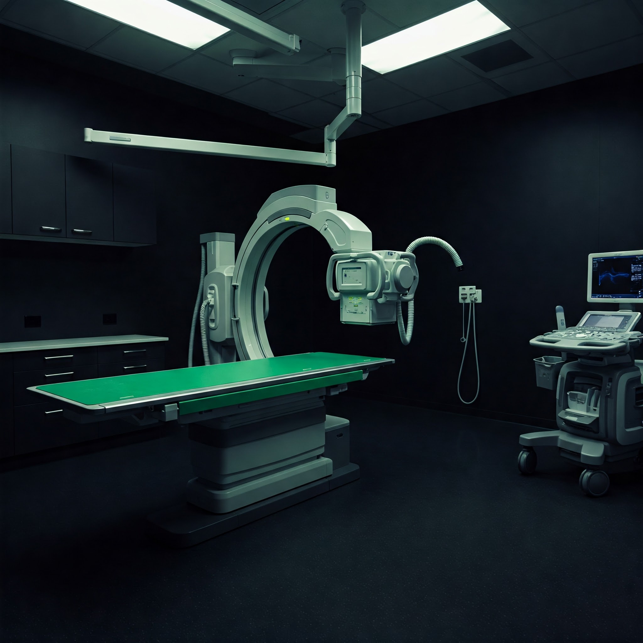
Advanced Sonography (Ultrasound)
Safe, radiation-free imaging using sound waves for detailed visualization of internal organs, tissues, blood flow, and fetal development.
- - Abdomen and Pelvis: Comprehensive evaluation of abdominal and pelvic organs.
- - KUB (Kidneys, Ureters, Bladder): Assessment for stones, infections, or other urinary tract issues.
- - Chest: Evaluation for pleural effusion or other chest conditions visible on ultrasound.
- - Follicular Monitoring: Tracking ovarian follicle development for fertility treatments.
- - Elastography: Advanced technique to assess tissue stiffness, often used for liver evaluation.
- - 1st Trimester Viability/Dating Scan: Confirms pregnancy, checks heartbeat, and determines gestational age.
- - Growth Scan: Monitors fetal growth and well-being in later pregnancy.
- - NT Scan (Nuchal Translucency): Early screening for chromosomal abnormalities (part of first-trimester screening).
- - Anomaly Scan (TIFFA): Detailed examination around 18-20 weeks to check fetal anatomy for abnormalities.
- - Colour Doppler Studies: Visualization of blood flow direction and velocity.
- - Carotid Doppler: Assesses blood flow in the neck arteries supplying the brain.
- - AV Fistula Doppler: Evaluation of arteriovenous fistulas, typically for dialysis patients.
- - Upper & Lower Limb Arterial/Venous Doppler: Checks for blockages, clots (DVT), or poor circulation in arms and legs.
- - Portal Doppler: Examines blood flow in the portal vein system (liver).
- - Renal Doppler: Evaluates blood flow to and within the kidneys.
- - Obstetrics Doppler: Assesses blood flow in the umbilical cord and fetal vessels.
- - Musculoskeletal (MSK) Ultrasound: Imaging of joints (Shoulder, Knee, Ankle), tendons, and muscles.
- - Sono-mammography (Breast Ultrasound): Detailed imaging of breast tissue, often complementary to mammography.
- - Neurosonography: Ultrasound imaging of the infant brain through the fontanelle.
- - B Scan (Ocular Ultrasound): Ultrasound imaging of the eye.
- - Inguino-scrotal Region: Examination of the groin and scrotum.
- - Local Swelling / Small Parts: Evaluation of lumps, bumps, or specific small areas.
- - Thyroid / Neck Ultrasound: Imaging of the thyroid gland and structures in the neck.
Digital X-ray
Fast and efficient imaging using minimal radiation, primarily for visualizing bones and certain conditions in the chest and abdomen.
- - Chest: Evaluation of lungs, heart, and chest wall.
- - Abdomen: Can help identify bowel issues, obstructions, or certain types of stones.
- - Spine: Imaging of cervical, thoracic, lumbar, or sacral spine.
- - Joints: Examination of various joints for arthritis, injury, or other conditions.
- - Extremities: X-rays of arms, legs, hands, and feet, typically for fractures or bone abnormalities.
- - Skull: Imaging of the bones of the head.
USG Guided Procedures
Utilizing ultrasound guidance for precise minimally invasive procedures:
- - Drainage: Guided drainage of Abscess, Ascites (abdominal fluid), and Pleural Effusion (chest fluid).
- - FNAC (Fine Needle Aspiration Cytology): Obtaining cell samples from lumps or organs for diagnosis.
- - Biopsy: Obtaining tissue samples under ultrasound guidance for detailed pathological analysis.
- (Availability may depend on specific case requirements)
Dr. Nemade and our team ensure clear communication throughout your procedure.
Advantages of Our Imaging Modalities
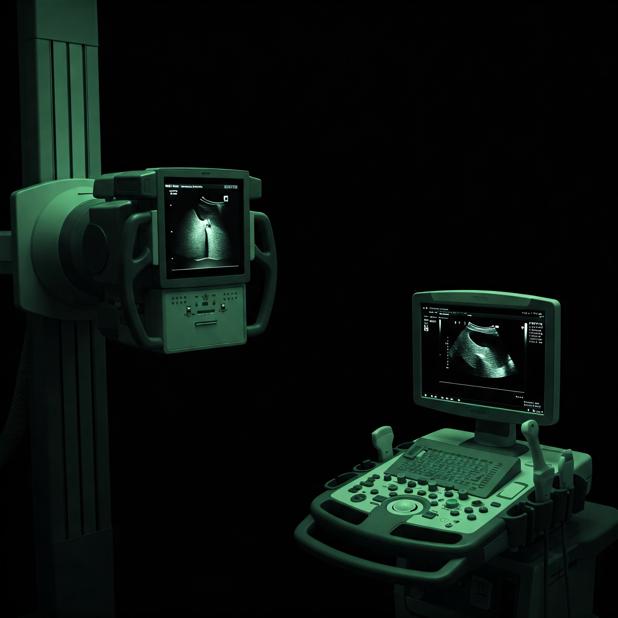
Why Choose Sonography?
Sonography offers several unique advantages:
- - Radiation-Free Safety: Ideal for all patients, including pregnant women, children, and for repeated examinations.
- - Real-Time Imaging: Allows visualization of movement, such as blood flow (Doppler) or fetal activity, providing dynamic information.
- - Excellent Soft Tissue Detail: Superior visualization of organs (liver, kidney), muscles, tendons, and blood vessels compared to X-ray.
- - Non-Invasive & Painless: Most ultrasound exams are comfortable and do not require needles or contrast agents.
- - Cost-Effective & Accessible: Generally more affordable and widely available than other advanced imaging like MRI.
- - No Claustrophobia Issues: An open procedure, making it suitable for patients uncomfortable in enclosed spaces.
The Role of Digital X-ray
Digital X-ray remains a cornerstone of diagnostics due to:
- - Speed and Efficiency: X-ray exams are very quick to perform, providing rapid diagnostic information, crucial in emergencies.
- - Superior Bone Imaging: Unmatched clarity for visualizing bone structures, fractures, dislocations, and joint abnormalities.
- - Wide Availability: A fundamental and readily accessible imaging technique found in most healthcare settings.
- - Cost-Effective: Generally less expensive than other imaging modalities like CT or MRI.
- - Supports Treatment Planning: Essential for guiding treatment decisions for bone injuries and certain lung/abdominal conditions.
- - Procedure Guidance: Can be used to guide certain medical procedures like injections or line placements.
A Note on Radiation Safety
We prioritize your safety. Our modern digital X-ray systems use the lowest necessary radiation dose (ALARA principle). The diagnostic benefits typically far outweigh the minimal risks associated with this low exposure.
Dr. Nemade helps determine the most appropriate imaging test for your specific clinical needs.
Preparing for Your Exam
Following preparation instructions carefully helps ensure the highest quality images for accurate diagnosis. General guidelines are below, but please follow any specific instructions provided by our staff or your doctor.
Sonography Preparation Details
Preparation varies by the type of ultrasound:
- - Pelvic (Transabdominal): Requires a full bladder. Drink approximately 1 litre (4-5 glasses) of water finishing 1 hour before your exam. Do not empty your bladder (except for heart failure patients or those advised restricted fluid intake).
- - Pelvic (Transvaginal): Usually performed with an empty bladder. You may be asked to empty your bladder just before the exam.
- - Other Ultrasounds (Breast, MSK, Doppler, Thyroid etc.): Typically no special preparation needed. Avoid lotions or powders on the area being examined.
- - Medications: Generally, continue taking prescribed medications as usual unless specifically advised otherwise.
- - Gall bladder: Usually requires fasting (no food or drink, except sips of water for medication) for 8-12 hours beforehand.
X-ray Preparation Details
Most X-rays require minimal preparation:
- - General: You may be asked to change into a gown and remove jewelry, eyeglasses, and any metal objects that could interfere with the images.
- - Chest / Bone / Abdominal X-rays: Typically no specific dietary preparation required unless instructed otherwise.
- - Contrast X-rays (if offered, e.g., Barium studies): These require specific instructions regarding fasting, diet, or bowel preparation. Follow these instructions precisely.
- - Pregnancy: Always inform the technologist if there is any possibility you might be pregnant before an X-ray exam.
Questions? If you are unsure about preparation instructions for your specific exam, please call us at +91 8956607770 / +91 9270460525.
Frequently Asked Questions
Find answers to common questions about our services and procedures.
Sonography (Ultrasound)
- - What is Sonography/Ultrasound? It uses high-frequency sound waves to create images of internal body structures.
- - Is it safe? Yes, it does not use ionizing radiation and is considered very safe, even during pregnancy.
- - Does it hurt? No, it's generally painless. You might feel slight pressure from the transducer (probe).
- - How long does it take? Most exams take between 10 to 15 minutes, depending on the area and complexity.
- - What is a Doppler Ultrasound? It's a special technique used to evaluate blood flow through arteries and veins.
- - Who performs the ultrasound? A trained sonographer performs the scan, which is then interpreted by Dr. Nemade.
- - When will I get the results? Dr. Nemade interprets the images, and a report is sent to your referring doctor, usually within 24-48 hours.
- - Do I need to bring anything? Bring your doctor's referral form, ID, and any relevant previous imaging reports.
Digital X-ray
- - What is an X-ray? It uses a small, controlled amount of ionizing radiation to create images of internal body structures, especially bones.
- - Is it safe? How much radiation? Modern digital X-rays use very low radiation doses. We follow the ALARA principle (As Low As Reasonably Achievable). The diagnostic benefit almost always outweighs the minimal risk.
- - Does it hurt? No, the X-ray procedure itself is painless.
- - How long does it take? The actual image capture takes seconds. Positioning might take a few minutes.
- - Who interprets the X-ray? The images are personally interpreted by our highly specialized radiologist, Dr. Nemade (MD Radiology).
- - Why might multiple images be needed? Different views (angles) are often required to fully visualize the area of interest.
- - What is the lead apron for? It's used to shield parts of the body not being imaged from unnecessary radiation exposure.
- - Can I get a copy of my images/report? Please discuss this with our staff or your referring doctor regarding the process.
Appointments & General Info
- - Do I need an appointment? Yes, appointments are generally required. Please call +91 8956607770 or use our contact details.
- - What should I bring? Your doctor's referral (if applicable), ID card, insurance details (if any), and any relevant prior medical reports or images.
- - What are your operating hours? Monday to Saturday: 10:00 AM - 3:00 PM and 5:00 PM - 7:00 PM. Sunday closed. (Appointments Required). Please call to confirm.
- - What payment methods are accepted? Pay by Cash, UPI.
- - What is your cancellation policy? Please inform us in advance if you need to reschedule or cancel.
- - Is the centre accessible? Yes, we have stretcher-compatible lifts and accessible facilities at Nashik Business Park.
About Dr. Nikita Nemade
Our clinic, Poorti Advanced Sonography Centre, is led by Dr. Nikita Nemade, a dedicated and experienced Consultant Radiologist.

Consultant Radiologist
Dr. Nemade is committed to providing accurate diagnostic imaging with a focus on patient care and clear communication. Her expertise covers a wide range of sonography and X-ray modalities.
M.B.B.S (K.E.M. Hospital, Mumbai), MD Radiology (Pune)
Contact Us & Location
Reach out to schedule your appointment with Dr. Nikita Nemade at Poorti Advanced Sonography Centre, ask questions, or find our centre located conveniently at Mumbai Naka, Nashik.
Our Location
Dr. Nikita Nemade (MD Radiology) - Poorti Advanced Sonography Centre
Shop no. 203, Nashik business park
Near Mahamarg bus stand, next to Saheba Hotel, Mumbai Naka
Nashik, Maharashtra
Phone:
+91 8956607770 / +91 9270460525
Email:
ultrasonodrnemade@gmail.com
Operating Hours:
Mon-Sat: 10:00 AM - 3:00 PM & 5:00 PM - 7:00 PM (Appointments Required)
Find us at Nashik Business Park, Mumbai Naka. Click the map for directions. Hours: Mon-Sat 10:00 AM - 3:00 PM & 5:00 PM - 7:00 PM (Appointments Required)